
Theranostics in nuclear medicine is a modern, mainstream approach that unites cancer diagnosis and treatment into a single, integrated system. It not only enables precise identification of tumors and their metastases but also allows targeted radionuclide therapy. Today, these methods are increasingly being included in international treatment protocols and offer patients a real chance to extend their lives and improve quality of life.
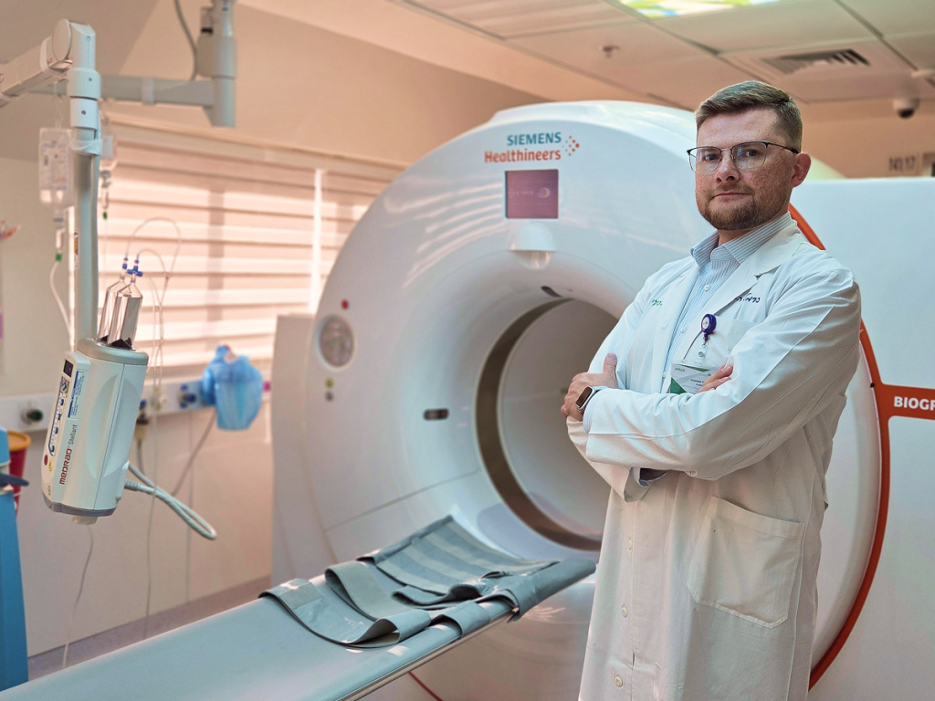
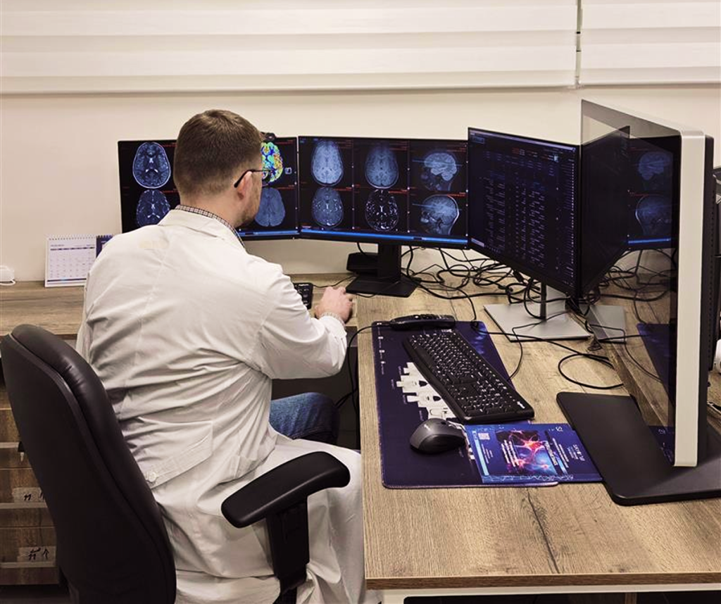
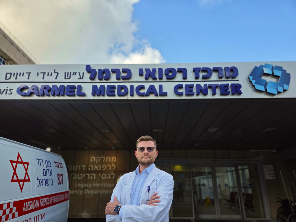
We discussed this with Dr. Konstantin Kenigsberg, Oncoradiologist and Nuclear Medicine Physician at Carmel Medical Center, and Israel’s representative at the European Society of Oncologic Imaging (ESOI). Dr. Kenigsberg explained why theranostics is seen as a major breakthrough in recent years and where the field is heading in the coming decade.
2digital: When we talk about theranostics in nuclear medicine, are we essentially describing a new approach to the combined diagnosis and treatment of cancer?
Dr. Kenigsberg: Yes and no. The term theranostics only came into common use relatively recently — in the 2010s. From that perspective, yes, it’s considered a new field. But if we look at it historically, the concept goes back much further. As early as the mid-20th century, doctors were treating thyroid tumors with radioactive iodine. In essence, that was already theranostics, just in a very limited form and not in the way we envision it today.
2digital: Then let’s start with the basics. What’s the essence of theranostics?
Dr. Kenigsberg: In nuclear medicine, the focus has traditionally been on diagnostics. The key lies in pairing an isotope with a molecule — together, this combination is called a tracer. The radiopharmaceutical must deliver the isotope precisely to the target — the tumor. That’s why researchers select special carrier compounds that ensure the isotope accumulates as selectively as possible in the tumor and its metastases. This allows us to “highlight” the tumor and identify its exact location.
It’s crucial to determine what stage the disease is at. Take prostate cancer, for example — the treatment strategy depends on how far it has spread. If the tumor is localized (confined to the prostate), surgery may be sufficient. But if the disease has spread throughout the body (metastases in lymph nodes, soft tissues, or organs), both the diagnostic and therapeutic approach must change entirely.
This is why whole-body scanning became necessary, and ideally, in a highly specific way, to detect all the “hot spots” where the radiopharmaceutical is pathologically accumulating. At the same time, we need to differentiate tumor uptake from physiological uptake in healthy tissues. Ideally, the drug should be absent — or present only in minimal amounts — in healthy tissues, and its distribution should be predictable and well understood.
2digital: How has nuclear medicine evolved from a diagnostic perspective?
Dr. Kenigsberg: In the mid-20th century, alongside isotopes and radiopharmaceuticals, scanners were also developing. Initially, these were planar scans — images were taken from the front and back of the patient, often full-body, or focused on specific regions like an arm. Then came SPECT — Single Photon Emission Computed Tomography. It captures gamma rays emitted by the isotope inside the body and creates a 3D image of the region of interest — though at quite a low spatial resolution.
Here’s the challenge: we get a 3D image, but we still need to understand the anatomical location of the pathology. Say we detect hot spots in the abdomen — they could be in the liver, intestines, vertebrae, or lymph nodes. To resolve this ambiguity, we need anatomical visualization.
That’s how hybrid scanners were developed — known as SPECT/CT (Single Photon Emission Computed Tomography combined with standard CT). One part of the machine provides the functional SPECT image based on isotope-labeled molecules, and the other offers anatomical context. By overlaying these two images, we can accurately pinpoint where the radiopharmaceutical is accumulating.
Later, PET — Positron Emission Tomography — was introduced. This is a more complex and more expensive technology, offering higher spatial resolution and the ability to use a broader range of tracers. Today, PET is the most in-demand imaging modality for diagnostic purposes.
2digital: Can you briefly summarize the advantages of theranostics?
Dr. Kenigsberg: The key advantage is that we can take virtually the same molecule used for effective diagnostics and “reconfigure” it for therapy. For example, when treating prostate cancer, we use a compound called PSMA — Prostate-Specific Membrane Antigen. This molecule is bound to a diagnostic isotope to create a tracer like [⁶⁸Ga]PSMA or [¹⁸F]PSMA. Once injected into the body, it allows us to visualize the prostate cancer and its metastases — for instance, in lymph nodes or bone marrow.
In certain cases, for patients with metastatic prostate cancer who are no longer responding to first- or second-line therapies, a theranostic option may be offered: we take the same PSMA molecule, but instead of [⁶⁸Ga] or [¹⁸F], we bind it to Lutetium-177 creating [¹⁷⁷Lu]PSMA. This is a different isotope — a beta emitter. Whereas the earlier isotopes emit radiation suitable for imaging, beta-emitters are therapeutic. Diagnostic radionuclides emit photons for visualization; therapeutic beta isotopes emit particles that deliver energy over very short distances — just a few millimeters. This damages the DNA of tumor cells and leads to their death, while minimizing harm to surrounding healthy tissue.
Such compounds are already included in clinical guidelines and are actively used in the treatment of metastatic prostate cancer, among other conditions.
Today, the concept is evolving further: researchers are developing agents based not only on beta emitters but also on alpha emitters. Alpha particles carry significantly more energy and have a high linear energy transfer (LET), making them more effective at destroying tumor cells while still sparing surrounding tissues. However, alpha-emitting radionuclides are extremely scarce: their production is limited, with only a handful of centers worldwide capable of producing them at clinically meaningful scale — and at very high cost.
Nonetheless, accumulating clinical evidence already suggests that therapies using alpha emitters may prove even more effective than the beta-based therapies now entering routine clinical use.
In addition to swapping out tracers in terms of the isotopes used — ones that enable decay and biological damage — we’re also seeing the emergence of new ligand molecules for diagnosing and treating different types of tumors. Until recently, theranostics was applied mainly to prostate cancer and neuroendocrine tumors — these are already FDA-approved and implemented globally, especially in high-income countries.
One of the recent breakthroughs that drew considerable attention is the development of the molecule FAPI — Fibroblast Activation Protein Inhibitor.
2digital: Tell us more about it.
Dr. Kenigsberg: FAPI is a new molecule used in diagnostics, and trials are already underway to assess its therapeutic and theranostic potential. Fibroblasts are connective tissue cells that form the body’s structural “scaffolding.” They’re abundant in the skin and extracellular matrix, producing collagen, elastin, and other proteins. Tumors also contain fibroblasts — but these differ somewhat from normal ones. Their role in the tumor microenvironment is to construct a protective matrix that helps the tumor evade the body’s immune system.
FAPI was specifically designed to bind to these activated fibroblasts within the tumor stroma. When combined with widely used molecular imaging isotopes like Fluorine-18 or Gallium-68, FAPI creates a highly effective radiopharmaceutical. Compared to most other tracers, FAPI compounds offer an excellent tumor-to-background uptake ratio — a high signal-to-noise contrast.
FAPI is especially useful in detecting tumors that don’t accumulate other commonly used tracers. For example, the most widely used radiopharmaceutical globally is [¹⁸F]FDG — a glucose analog. It accumulates in tissues with high glucose metabolism. But some tumor types either don’t absorb FDG at all or show very weak uptake — such as primary liver cancer, certain kidney cancers, sarcomas, pancreatic tumors, and malignancies of the gastrointestinal tract, among others.
This is where FAPI shows real promise: available data suggest it achieves consistently high uptake in many of these malignancies. Researchers are now actively investigating its potential in both diagnosis and therapy across a wide spectrum of cancers — and so far, the results look highly encouraging.
2digital: Would you say that modern theranostics is a breakthrough in cancer care?
Dr. Kenigsberg: Theranostics is definitely a breakthrough — though not an overnight one. It’s more of a work in progress. The foundations were laid back in the mid-20th century, but the field has gained real momentum in just the past decade — thanks to rapid advances in imaging technology and radiopharmacology.
What we’re witnessing now is truly personalized medicine: treatment tailored to the specific type of cancer an individual patient has. We begin with diagnostic imaging to map out where the tumors are located in the body, and then introduce a radiopharmaceutical with a different isotope that selectively destroys tumor DNA using targeted radiation. After treatment, we return to diagnostic tracers to assess how effective the therapy was. This is personalized care — highly specific, highly precise.
Of course, like everything in medicine, it’s not without its limitations — we know that there are no 100% guarantees. But the results we’re seeing are genuinely promising and encouraging.
And we can’t talk about modern medicine without mentioning AI. AI is being integrated into theranostics on multiple fronts. For example, it allows us to significantly reduce radiation exposure during diagnostics through smart image reconstruction, and it also helps tailor the exact dose of radiopharmaceutical for therapy — a critical part of nuclear medicine known as dosimetry.
Sometimes, AI enables us to see things the human eye can’t. Through radiomics and texture analysis of imaging data — another highly promising direction — AI can help predict how a disease might progress, even before therapy begins. We already have established criteria for determining who qualifies for therapy and who doesn’t. But AI has the potential to expand these boundaries — to predict whether a patient is likely to benefit from treatment, and what their chances of a positive outcome might be, by analyzing “invisible” features in the images that correlate with the biological properties of their tumor.
Radiomics and texture analysis are actively being researched in both radiology and nuclear medicine, although they’re not yet part of standard clinical protocols. I believe AI will give this area a real boost and help unlock its full technological potential.
Lastly, it’s important to highlight that theranostics is primarily used as a second- or third-line therapy — and it truly improves both survival and quality of life. We’re often talking about advanced, metastatic cancers, where the patient has very few treatment options left — and frankly, a poor prognosis.
Theranostics offers a way to change that trajectory.
In many cases, it becomes the “lifeline” patients and doctors had been hoping for — even knowing that the odds were slim. And now, that chance — cautiously speaking — is becoming real. It may not always lead to a cure (although there are cases when it does), but it can meaningfully extend life and improve its quality.
2digital: In your view, where is theranostics headed in the next decade?
Dr. Kenigsberg: First, diagnostics will become more precise, as technological advances allow us to process much larger volumes of data — faster and more accurately than ever before. Automated segmentation, complex post-processing and image analysis, including radiomics and texture analysis, will make oncology diagnostics truly personalized.
Second, we’ll be able to develop new molecules more quickly, and the arsenal of radiopharmaceuticals available for clinical use will grow. Today, it typically takes around 10 years to bring a radiopharmaceutical from concept to clinical application. I’m confident that over the next decade, this timeline will be significantly shortened — and we’ll have many more options for both diagnosis and treatment of various malignancies.
Third, the treatment process itself will improve. We’ll scan, transmit, and analyze data faster and collect higher-quality information — both of which will enhance the standard of care. And hopefully, it will also increase accessibility: making these cutting-edge methods — currently concentrated in high-income countries — more available across the globe.